Fetus Growth Evaluation Tool
At any gestation, if the ultrasound fetal measurements of each parameter are not in agreement, the reason for this difference should be evaluated.
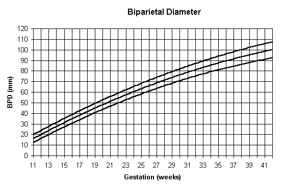
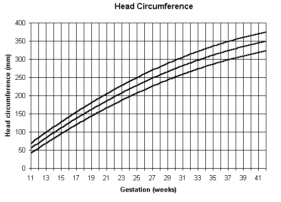
BIPARIETAL DIAMETER AND HEAD CIRCUMFERENCE
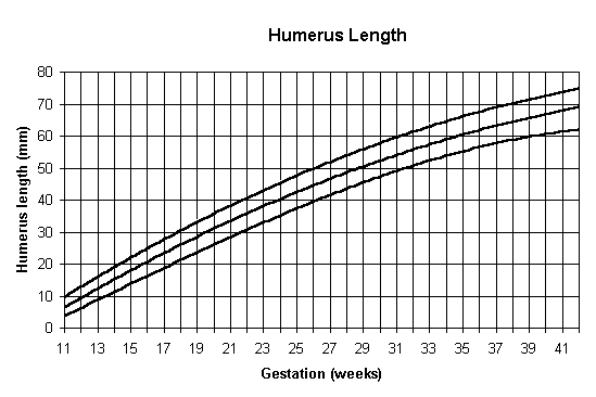
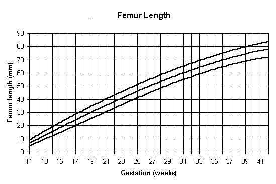
FEMUR AND HUMERUS LENGTH
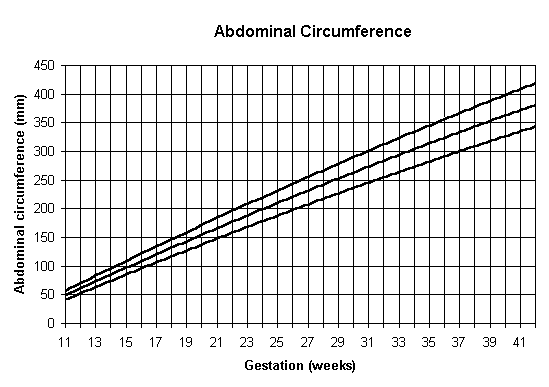
ABDOMINAL CIRCUMFERENCE
Include two fetal measurements (usually biparietal diameter (BPD) and femur length (FL)) plus a consideration of additional parameters such as head circumference (HC), occipitofrontal diameter (OFD), abdominal circumference (AC) and humerus length (HL).
The wide normal range of BPD in late pregnancy must be appreciated. It is not expected that BPD be used to assess gestation late in pregnancy. The values from 33 weeks are intended to predict the growth in fetal head size from a known gestation.
The most common are:
Gestational Sac:
The first element to be measurable is the gestation sac of the early pregnancy. The gestational sac is measured in three dimensions, and the average, the Mean Sac Diameter (MSD) used for estimating gestational age. It is useful between 5 and 8 menstrual weeks with accuracy of +/- 3 days . As a rough rule of thumb, the MSD + 30 = Menstrual Age in days.
Embryonic Crown-Rump Length:
The length of the embryo on the longest axis (excluding the yolk sac) constitutes the crown-rump length. This is among the best documented parameters to date the embryo, with accuracy of +/- 3-5 days. As a rough rule of thumb, the CRL + 6.5 = Menstrual Age in Weeks.
Biparietal diameter (BPD):
The transverse width of the head at it's widest. We measure from the the leading edge to leading edge of the bones, because this leading interface is most distinct. Since the head is oval, the error induced by small errors in positioning is small, making for a repeatable, robust measure.
Head size is determined largely by brain growth which is relatively independent of nutritional (maternal/placental insufficiency) growth retarding processes, and head growth is often relatively "spared" in such growth retardation.
The BPD best used after 12 weeks.
Accuracy is +/- 1.1 week 14 - 20 weeks,
+/- 1.6 weeks 20 - 26 weeks,
+/- 2.4 week 26 - 30 weeks,
and +/- 3 - 4 weeks after 30 weeks.
Femur Length:
The femur length is a repeatable measurement with accuracy similar to the BPD. It is effected by skeletal dysplasias, but since these are rare, it is a reliable measurement which confirms measurements of the head. It is best measured after 14 weeks.
It is common to make at least 2 and often 4-5 measurements to estimate gestational age. In most cases, BPD, Head Circumference, Femur length, and Abdominal circumference are used.
Since all of these measurements are strongly related to gestational age, it is not
usually important how they are combined. Most modern ultrasound machines include
computerized biometric analysis programs used to easily calculate your Estimated
Due Date or EDD or EDC.
Ultrasound Fetal Measurement Standards Chart
NOTE: +/- Standard deviations shown in (brackets). Measurements are for completed weeks.
| Gestation (weeks) |
BPD (mm) |
OFD (mm) |
Head circumference (mm) |
Abdominal circumference (mm) |
Femur (mm) |
Humerus (mm) |
Gestation (weeks) |
| 11 | 16 (2.0) | 21 (2.0) | 59 (15) | 52 (10) | 8 (2.0) | 8 (3.0) | 11 |
| 12 | 20 (4.0) | 24 (2.0) | 70 (15) | 63 (10) | 10 (2.5) | 9 (2.0) | 12 |
| 13 | 24 (4.0) | 29 (3.0) | 84 (15) | 74 (10) | 11 (2.5) | 11 (3.0) | 13 |
| 14 | 28 (4.0) | 34 (3.0) | 96 (15) | 84 (10) | 15 (3.0) | 14 (4.0) | 14 |
| 15 | 31 (4.0) | 38 (3.0) | 108 (15) | 96 (10) | 17 (3.5) | 17 (5.5) | 15 |
| 16 | 36 (5.0) | 46 (3.0) | 128 (15) | 106 (10) | 22 (4.0) | 21 (4.0) | 16 |
| 17 | 39 (5.0) | 50 (3.0) | 141 (15) | 120 (15) | 25 (4.0) | 25 (5.0) | 17 |
| 18 | 42 (4.0) | 54 (3.5) | 151 (20) | 131 (15) | 28 (5.0) | 27 (5.5) | 18 |
| 19 | 45 (5.0) | 57 (3.5) | 160 (20) | 140 (15) | 30 (5.0) | 29 (5.0) | 19 |
| 20 | 47 (4.0) | 61 (3.5) | 170 (20) | 151 (15) | 32 (6.0) | 31 (5.0) | 20 |
| 21 | 49 (4.0) | 63 (4.0) | 176 (20) | 164 (20) | 34 (6.0) | 32 (6.0) | 21 |
| 22 | 52 (5.0) | 68 (3.5) | 188 (20) | 176 (20) | 37 (5.0) | 35 (6.0) | 22 |
| 23 | 57 (5.0) | 76 (4.0) | 210 (20) | 186 (20) | 43 (5.0) | 38 (4.0) | 23 |
| 24 | 60 (6.0) | 79 (4.0) | 220 (20) | 201 (20) | 45 (4.0) | 40 (6.0) | 24 |
| 25 | 64 (6.0) | 82 (4.5) | 231 (20) | 212 (20) | 48 (5.0) | 43 (5.0) | 25 |
| 26 | 67 (4.0) | 84 (4.5) | 238 (20) | 223 (25) | 49 (5.0) | 44 (4.0) | 26 |
| 27 | 68 (5.0) | 86 (4.5) | 250 (20) | 230 (25) | 50 (5.0) | 47 (4.0) | 27 |
| 28 | 72 (4.0) | 95 (5.0) | 263 (20) | 242 (25) | 54 (4.0) | 50 (5.0) | 28 |
| 29 | 75 (4.0) | 97 (5.5) | 269 (25) | 259 (25) | 55 (5.5) | 51 (5.0) | 29 |
| 30 | 76 (4.0) | 98 (5.5) | 274 (25) | 262 (25) | 58 (6.0) | 52 (5.0) | 30 |
| 31 | 80 (6.0) | 101 (5.0) | 284 (25) | 272 (30) | 59 (5.5) | 54 (5.0) | 31 |
| 32 | 81 (4.0) | 102 (5.0) | 288 (25) | 283 (30) | 62 (6.0) | 56 (5.0) | 32 |
| 33 | 84 (6.0) | 107 (5.5) | 300 (25) | 294 (30) | 65 (4.0) | 57 (6.0) | 33 |
| 34 | 86 (6.0) | 108 (5.5) | 305 (25) | 305 (30) | 66 (4.0) | 59 (5.5) | 34 |
| 35 | 88 (6.5) | 109 (5.5) | 310 (25) | 315 (30) | 67 (6.0) | 60 (6.0) | 35 |
| 36 | 90 (6.0) | 112 (5.5) | 317 (25) | 325 (35) | 69 (6.0) | 62 (5.0) | 36 |
| 37 | 92 (6.5) | 113 (6.0) | 321 (25) | 333 (35) | 72 (5.0) | 63 (6.0) | 37 |
| 38 | 93 (6.0) | 116 (5.5) | 328 (25) | 342 (35) | 73 (5.5) | 64 (6.0) | 38 |
| 39 | 95 (8.0) | 119 (6.0) | 336 (25) | 356 (35) | 75 (6.0) | 65 (5.5) | 39 |
| 40 | 96 (8.0) | 120 (6.0) | 340 (25) | 362 (35) | 76 (4.0) | 66 (6.0) | 40 |
| 41 | 98 (8.0) | 122 (6.0) | 344 (25) | 367 (35) | 77 (5.0) | 68 (6.0) | 41 |
How many scans should I have?
As a practical matter, ultrasound scanning has proven to be so popular with patients and also their obstetricians, that almost everyone receiving regular prenatal care ends up with at least one scan anyway. The total number of scans will vary depending on whether a previous scan has detected certain abnormalities that require follow-up assessment. There is no hard and fast rule for the number of scans you should have during pregnancy. In some countries, sonograms are performed just twice during pregnancy. Once at 16 - 18 weeks to assess fetal abnormalities, and again at 32 - 34 weeks to assess age and well-being. It is now recommended that all pregnant women have a dating scan in the first trimester - ideally at 10 to 13 weeks of pregnancy - to confirm your dates. This is especially important if you are going to have any screening tests for Down's syndrome, as knowing the exact dates makes sure your result is accurate. Most hospitals offer a scan in the second trimester at about 20 weeks (the anomaly scan) to check that your baby is developing normally.